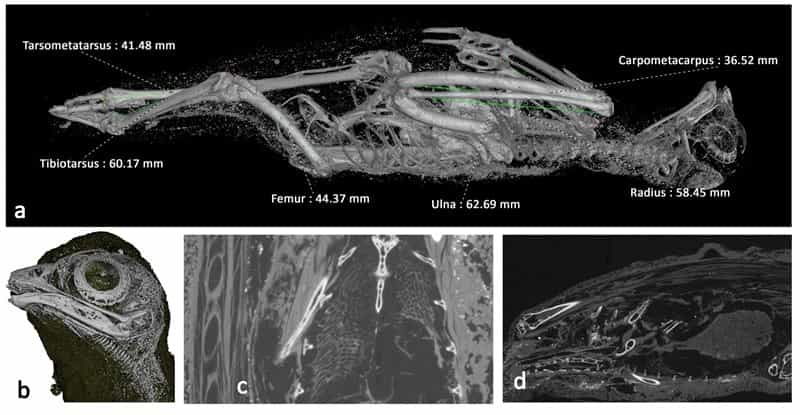
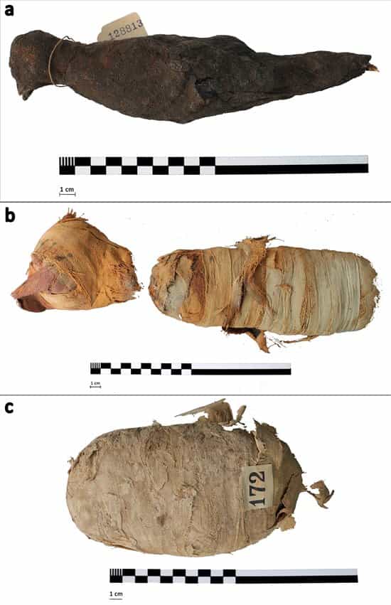
A team of researchers from Swansea University has used X-ray microtomography technology to obtain high-resolution, 3D images of three ancient Egyptian animal mummies: a cat, a bird, and a snake, all mummified more than 2,000 years ago.
The innovative technology has provided unprecedented detail without the need to physically open the mummies, revealing much about their content and purpose.
The animals scanned were a domestic cat, an Egyptian cobra, and a common kestrel, all of which were sacred to the ancient Egyptians. The images indicate that they were likely votive offerings to the gods, a common practice in ancient Egypt.
It is estimated that the ancient Egyptians mummified up to 70 million animals, mostly as offerings to the gods, but also with their pets for the afterlife and with cattle as food.
X-ray microtomography combines the benefits of tomography and radiography, providing 3D images with a resolution 100 times greater than previous techniques. The high resolution allows for details such as bones and teeth to be seen, leading to new discoveries such as the cat’s age and cause of death.
The study was conducted by a multidisciplinary team led by Professor Richard Johnson from Swansea University, in collaboration with the Egypt Center, a museum of Egyptian antiquities in Wales, and universities of Leicester and Cardiff. According to Dr. Carolyn Graves-Brown from the Egypt Center, this collaboration between engineers, archaeologists, biologists, and Egyptologists highlights the value of researchers from different fields working together to better understand the secrets of ancient Egypt.
Source: Abel G.M., National Geographic


